Essentials of Anatomy and Physiology for Nursing Practice
Second Edition
by Neal Cook, Andrea Shepherd and Jennifer Boore
Student Resources
Answers to Revise Questions
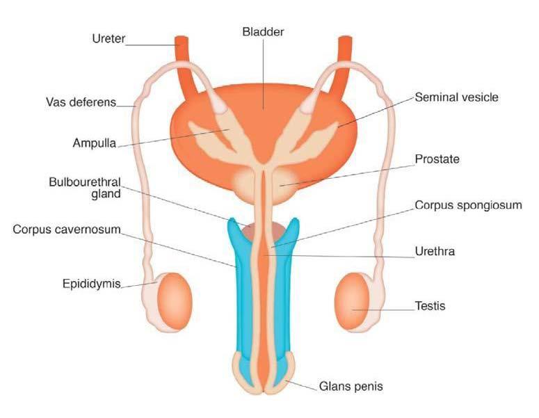
Label the diagram of the male reproductive system.
Ans:
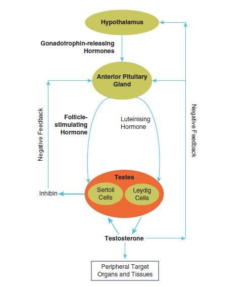
Draw a diagram to show the endocrine regulation of the male sex organs.
Ans:
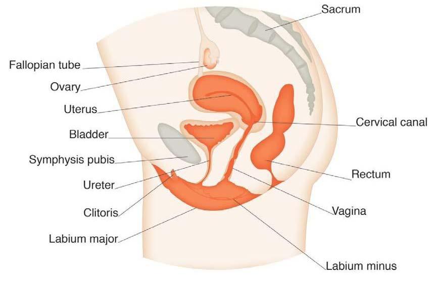
Label the diagram of the female reproductive system.
Ans:
Outline the endocrine changes during the menstrual cycle and the effects on the endometrium.
Ans:
- Pituitary hormones: FSH and LH reach peak at about mid-point of the cycle, stimulating development of an ovarian follicle. Ova released, the follicle becomes the corpus luteum.
- Ovarian hormones: Ovarian follicle releases oestrogen which slowly rises and peaks just before mid-point. Corpus luteum releases progesterone which readies the uterus for pregnancy, and then release falls to the end of cycle.
- Endometrium: Follicular phase as epithelium proliferates; in the luteal phase it continues to develop. As ovarian hormones fall to the lowest point, epithelium is shed through menstruation.
Outline the stages of the sexual response.
Ans:
- Excitement/arousal: dilation of blood vessels. P and BP up
Male->growth of penis, female->swelling of external genitalia.
- Plateau: changes above continue
Penis 50% larger, breast swells by 25%.
- Orgasm: contractions in pelvic floor and genital muscles->orgasm
Male->ejaculation, female->intense/pleasurable release of tension.
- Resolution: returns to normal
Recovery for men – minutes to hours, female slowly returns to normal, can orgasm again quickly.
Briefly describe the maternal changes during pregnancy.
Ans:
- Endocrine system:
- Progesterone and oestrogen levels rise.
- Placenta takes over secretion of oestrogen, human chorionic gonadotrophin (hCG), progesterone.
- Prolactin from pituitary prepares breasts for breast feeding.
- Parathyroid hormone ↑ -> levels of calcium ↑.
- Adrenal hormones (cortisol and aldosterone).
- Human placental lactogen (hPL) stimulates fat metabolism.
- Immune system: baby susceptible to immune attack. Normally maternal tolerance prevents this, the placenta acts as a barrier between the foetus and mother’s immune system.
- Physical changes:
- Increase in weight due to growth of the foetus and placenta, amniotic fluid, increased breast size and increase in water and fat retention.
- Changes in shape and posture of the woman’s body (Figure 16.21) ->change in centre of mass ->alterations in posture, gait, balance and increased risk of falls.
- Oestrogen and relaxin ->remodelling of soft tissues, ligaments and cartilage.
- Sacroiliac and symphysis pubis joints stretch and widen.
- Cardiovascular adaptation: plasma and blood volume increases, cardiac output ↑.
- BP falls (weeks 12–26), returns to pre-pregnancy levels by week 36.
- Increased erythropoietin secretion increases red blood cell numbers.
- Homeostasis: increased nutrients for baby and mother.
- Weight gain of about 20–30 lb (9.1 to 13.6 kg).
Additional protein for development of muscular tissues of the uterus and breast development for lactation and breast feeding.
What changes occur in the woman following delivery?
Ans:
- Returning to non-pregnant state: puerperium (6 weeks post-delivery the body returns to normal).
- Vaginal discharge:
- Lochia rubra (red): three to five days.
- Lochia serosa (brownish/pink): up to ten days.
- Lochia alba (white/yellow): 2nd through 3rd to 6th week.
- Infant feeding and nurturing:
- Milk ‘comes in’ 2nd or 3rd day.
- First colostrum with antibodies, then milk hormones involved.
- Prolactin – stimulates milk production.
Oxytocin – contraction of lactiferous ducts, squeezes milk out of the breast.