Essentials of Anatomy and Physiology for Nursing Practice
Student Resources
Answers to Revise Questions
What are the major functions of the nervous system?
Ans: The major functions of the nervous system are to maintain homeostasis by:
- receiving information and transmitting it to the central nervous system;
- integrating and analysing the different sources of information;
- making decisions;
- sending instructions to the muscles and glands of the body to:
- carry out voluntary movement;
- influence endocrine function (see next section);
- regulate unconscious activities through the autonomic nervous system.
Describe the divisions of the nervous system and their functions. Support this with a diagram of the structural divisions of the nervous system.
Ans: The central nervous system (CNS) is made up of the brain and spinal cord, which act as the control centre for many of the parameters that are controlled through homeostasis. The sensory division of the nervous system consists of the special senses and the general sensory receptors and nerves, which carry information to the CNS.
The motor division of the nervous system is made up of two parts:
- The somatic or voluntary nervous system, which causes movement of the skeletal muscle.
- The autonomic nervous system, which regulates involuntary activities. This is also in two parts:
- sympathetic: activated with ‘fright, flight or fight’;
- parasympathetic: activated under relaxed quiet conditions.
Describe how nerve impulses are transmitted.
Ans: Neurons carry information by transmitting nerve impulses along the axon in a way that is comparable to electrical charges. This is known as an action potential. The impulse passes from neuron to neuron at a synapse or neuron to muscle fibre at a neuromuscular junction by chemical or electrical transmission.
Neurons have two key properties:
- Irritability: this means that a stimulus initiates a nerve impulse. The stimulus may be external to the body (e.g., touch, light) or inside the body (e.g., concentration of CO2, temperature or thought).
- Conductivity: this means the ability to transmit an impulse.
Neurons may be myelinated or unmyelinated.
- Myelinated: the neurons of peripheral nerves and large axons are surrounded by a fatty sheath of myelin formed by Schwann cells or oligodendrocytes wrapped round and round the axon. Small spaces between the Schwann cells permit nerve impulses to ‘jump’ from space to space and travel rapidly along the nerve fibre.
- Unmyelinated: several of these neurons are embedded within a single Schwann cell. Nerve impulses are transmitted much more slowly than in myelinated neurons.
Describe in outline the major structural elements of the CNS and their functions.
Ans: The CNS consists of the brain and spinal cord and is composed of neurons and supporting tissue. They are made up of grey matter and white matter:
- Grey matter consists of the neuron cell bodies and is organised around the outer surface of the brain (the cerebral cortex) with clumps of grey matter in the middle of the brain, and in the centre of the spinal cord.
- White matter consists of myelinated nerve fibres passing between different parts of the brain and down towards the spinal cord or to the peripheral nervous system. This is in the inner parts of the brain but surrounds the grey matter of the spinal cord.
The nerve fibres are grouped in tracts or pathways. Two major tracts are:
- The corpus callosum, which joins the two sides of the brain.
- The internal capsule, which consists of both sensory and motor fibres which carry information to and from the cerebral cortex. (The internal capsule is a common site for a stroke which then results in damage to both sensory and motor pathways resulting in loss of sensation and loss of movement in the side of the body affected.)
Structurally, the brain is divided into:
The Forebrain: The largest section of the brain. It is associated with the control of body temperature, reproduction, eating, sleeping, cognition and emotional responses. It consists of the diencephalon (composed of the thalamus, hypothalamus and epithalamus) and the cerebrum (consisting of the cerebral cortex (grey matter) and underlying white matter, basal nuclei and limbic system).
The cerebral cortex is divided into lobes:
Frontal: regulates movement, cognition and personality.
Parietal: processes sensory information such as touch, vibration, proprioception, stereognosis and spatial perception. Also concerned with calculations, writing and reading.
Occipital: concerned with visual stimuli, recognising, interpreting and finally memorising objects.
Temporal: processes special senses (gustation, olfaction, audition), has a role in learning, memory, visual recognition and emotional actions. Processes interpretation of language.
Insular: thought to be involved in thermosensation, nociception, somatosensation, viscerosensation and gustation. Also part of the limbic system.
The Midbrain: Composed of tracts of nerve fibres and a number of nuclei of cranial nerves. Includes the substantia nigra which works with the basal nuclei to regulate movement through inhibiting the neurotransmitter/hormone dopamine. The cranial nerves responsible for eye movement originate in the midbrain, as do nuclei for the response to auditory and visual stimuli, such as turning your head to respond to a sound or visual stimulus. Corticospinal tracts pass through the midbrain on their way to the medulla, and reticulospinal tracts associated with the experience of pain are present.
The Hindbrain: Consists of the pons (acts as a relay centre, and regulates breathing through the pneumotaxic and apneustic centres), the medulla oblongata (acts as a relay centre, some corticospinal tracts decussate here, contains the cardiac, respiratory, vasomotor and reflex centres) and the cerebellum (coordinates the muscles of the body and regulates muscle tone and posture; also involved in cognition).
- Ascending (afferent) neurons carry information up the spinal cord to the brain for processing.
- Descending (efferent) neurons carry instructions down the spinal cord from the brain to the muscles and glands of the body.
- Interneurons (association neurons) act as connections between descending and ascending neurons.
The spinal cord is an extension of the brain from the medulla with two main enlargements. The cervical enlargement (through the brachial plexus) provides neurons for the arms, and the lumbar enlargement (through the lumbosacral plexus) provides neurons for the legs. Below this, the cord narrows into a conical shape, the conus medullaris. Nerves resembling a horse’s tail (called the cauda equina) extend further down to the sacrum.
Spinal grey matter is set out in an H shape and is divided into ‘horns’; anterior, posterior and lateral. Spinal white matter surrounds the grey matter and is structured into three columns, the anterior, posterior and lateral columns. They are then subdivided into tracts, distinct sets of fibres going to or from the same place. The name of the tract indicates its path; for example, the corticospinal tract carries information from the cortex to the spinal cord.
How many cranial nerves are there?
Ans: 12
What is the 5th cranial nerve called and what are its characteristics?
Ans: 5th or trigeminal nerve: mixed nerve innervating face. Trigeminal neuralgia causes pain along the route of the trigeminal nerve affecting the face and eye.
Which cranial nerve innervates the organs of the thorax and abdomen?
Ans: 10th or vagus nerve: innervates organs of the thorax and abdomen. Major nerve comprising the cranial end of the parasympathetic nervous system.
What happens to motor nerves as they pass through the medulla oblongata? Where does this happen for sensory nerves? What is the significance of this?
Ans: The nerve tracts from the motor cortex cross over (decussate) in the medulla oblongata so that the right side of the body is controlled by the left side of the brain and vice versa. The sensory nerve tracts cross over either in the medulla oblongata or in the spinal cord; thus, sensation from the left side of the body is received in the sensory cortex of the right side of the brain.
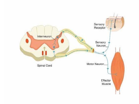
What is a reflex arc? Illustrate this in a diagram
Ans: A reflex arc is when the nerve impulse is transmitted rapidly from the sensory neuron through the connector neuron to the motor neuron without going through the brain. Therefore, the reflex arc is an important protective mechanism, as it permits a more rapid response than if information had to go the brain and back.
Briefly explain how information is transmitted to the central nervous system and represented in the cerebral cortex.
Ans: Information is carried by the sensory receptors into the spinal cord and up sensory pathways to the brain.
Sensory:
The sensory homunculus shows the representation of general sensory information in the sensory area of the cerebral cortex. Those areas of the body which have a high concentration of sensory receptors (e.g. the lips) are represented by a larger area of cortex than those with few receptors (e.g. the back).
Motor:
The axon of the motor neuron passes through the internal capsule, decussates in the medulla oblongata, and passes in a motor nerve tract to the spinal cord. In the spinal cord it synapses with the motor cell body. Note: One motor nerve passes all the way from the cerebral cortex to the motor cell body in the spinal cord. The motor pathway from the spinal cord to the periphery is known as the final common pathway, as it is the final nerve fibre on which all nerve impulses to that part of the body end.
What activities is the autonomic nervous system concerned with? What are the effector organs for this system? How is the autonomic nervous system structured, and what are the functions of its divisions?
Ans: The autonomic nervous system is concerned with control of involuntary activity of the body. The effector organs are as follows:
- Smooth muscle.
- Cardiac muscle.
- Glands.
The autonomic nervous system consists of two branches: Sympathetic and Parasympathetic Nervous Systems, which normally act together to regulate the activity of organs.
Sympathetic nervous system
This part of the autonomic nervous system derives from the thoracic and lumbar sections of the spinal cord. The cell bodies of the nerve fibres going to the individual organs are in a chain of ganglia, which run alongside the spinal column, with some in larger ganglia within the abdominal cavity.
As the effect of the sympathetic nervous system is to prepare the body for action, it activates those organs which enable increased activity (e.g. the heart and respiratory system) while, at the same time, it inhibits the activity of those organs (such as the gut) which have no active part to play in sudden activity.
Parasympathetic nervous system
This part of the autonomic nervous system derives from the cranial and the sacral ends of the CNS. Four of the cranial nerves contribute to this, the major one being the vagus nerve. The cell bodies of the nerve fibres are in the wall of the organ being supplied or in a ganglion within the abdominal cavity.
As the effect of the parasympathetic nervous system is to calm down the activities of the body and allow quiet restoration, in general it has an effect opposite to that of the sympathetic nervous system, slowing the heart and respiratory rates. It promotes the digestion and absorption of food.
Where is CSF formed and reabsorbed in the brain? How many fluid-filled spaces are there filled with CSF? Why do we have CSF?
Ans: Fluid (CSF) is formed in the ventricles of the brain by the choroid plexus. It circulates in the central canal of the spinal cord and around the brain and spinal cord. It is reabsorbed in the arachnoid villi.
The CSF surrounds and cushions the brain and spinal cord by maintaining a constant pressure of the fluid. It supplies nutrients and removes waste products from the nerve cells.
What are the functions of the meninges surrounding the central nervous system? Describe the three layers of the meninges.
Ans: Provide protection for the CNS.
- The dura mater: the thickest double layer. The outer layer is attached to the bone with the inner layer protecting the brain and forming the two structures separating the two hemispheres of the cerebral cortex and separating the cerebrum and the cerebellum. It encloses the spinal cord and the nerves continuing on down within the spinal column.
- The subdural space (a potential space) between the dura and the arachnoid mater.
- The arachnoid mater: is a fine membrane passing over the convolutions of the brain and envelops the spinal cord.
- The subarachnoid space contains the CSF, and separates the arachnoid and pia mater.
- The pia mater: is fine connective tissue adherent to the brain and spinal cord and dipping into each fissure. It contains fine blood vessels.
Briefly describe how blood is supplied to the brain.
Ans: The blood supply to the brain is arranged to enable the blood supply to be maintained even when one of the main supplying arteries is damaged. Blood from the two internal carotid arteries and the basilar artery (from the two vertebral arteries) enters the Circle of Willis, which allows the supply to be evened out to provide blood flow to the arteries supplying the different parts of the brain.